9
The extremities: hands and feet
The bones of the extremities (hands and feet), often generically termed ‘metapodials’ are numerous, some with confusingly similar morphologies. This chapter provides a generalised discussion of both fore and hind limb elements as a starting point. Two bones, the astragalus (talus) and calcaneus are discussed in detail, since both are found in all species, are easy to identify, and are commonly recovered intact from archaeological contexts. The remaining bones are discussed more generally. Photos of articulated extremities of each species are included to augment this discussion. The combination of text and photos provides a starting point that should permit you to at least identify a bone as coming from the hand or foot, and hopefully enable you to identify its type (e.g. metatarsal, phalanx). As you are by now no doubt aware, finer identification to species necessitates a good comparative collection and consultation with a specialist.
‘Hands’ versus ‘feet’
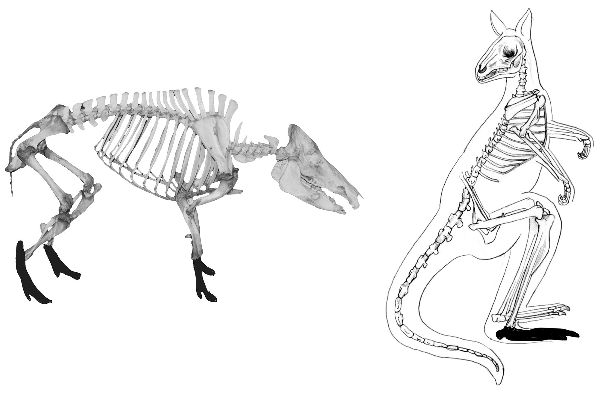
Many of the major bones of the extremities of the major quadrupeds are virtually indistinguishable, while there are greater differences among bipeds (in this manual, humans and macropods). The major bones of the fore leg (hand) are the carpals (wrist), metacarpals (knuckles) and phalanges (fingers) (Figure 9.1). The major bones of the hind leg are the calcaneus, astragalus, tarsals (all three make up the ankle joint), metatarsals and phalanges (toes) (Figure 9.2). From an evolutionary perspective, there is a successive reduction in the number of bones, beginning with humans. For example, the human foot has 26 bones (and there are 27 in the human hand), and horses have 12 foot bones.
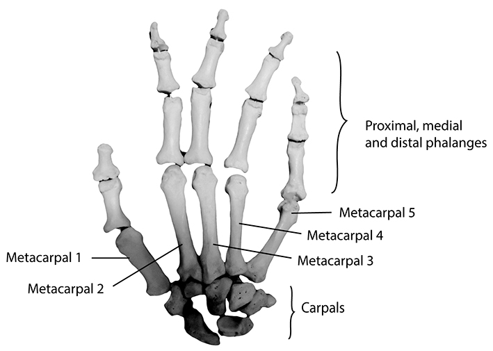
Figure 9.1: Human hand with major bones labelled.
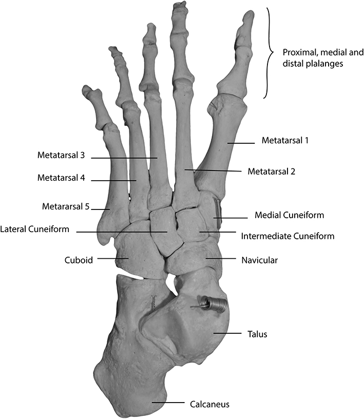
Figure 9.2: Human foot with major bones labelled.
Types of bones in the extremities
Carpals are the small, irregularly shaped bones that make up the fore limb ankle joint (or wrist in bipeds). The number of carpals varies by species, ranging from seven in humans to just two in cattle and sheep. The name of each varies according to position and whether the nomenclature followed is for humans or animals (see Hillson 1992).
Tarsals are the hind limb (ankle) equivalent to carpals, and they too vary in shape, number and morphology. The maximum number of tarsal bones is seven (found in most species) to two (found in cattle and sheep). The astragalus (talus) and calcaneus are the largest, and most easily recognisable of the tarsal bones, and are discussed and depicted in greater detail below (Figures 9.8–9.9).
The astragalus is one of two main bones in the ankle joint (Figure 9.3). It has two main articular facets, the superior, which is either saddle or pulley-shaped, for articulation with the distal tibia, and a second inferiorly, which varies in morphology, for articulation with the central tarsal bone.
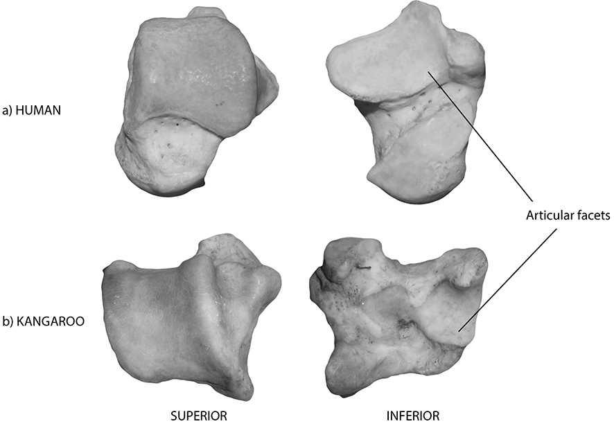
Figure 9.3: Astragali with the articular facets labelled in the superior and inferior views; (a) human, (b) kangaroo.
The calcaneus (the heel bone) is easy to recognise due to its distinctive morphology, which includes a square to triangular-shaped distal end with several articular facets (Figure 9.4). Proximally, the calcaneus has a swollen, roughened head (1), to which the Achilles tendon is attached. The shaft (2) varies in length and thickness, and the distal articulation contains a series of smaller articular surfaces for joints with the astragalus and central tarsal bones. There is a roughened protrusion on the medial size of the distal articulation, the sustentaculum tali (3), which forms the main articulation with the astragalus.
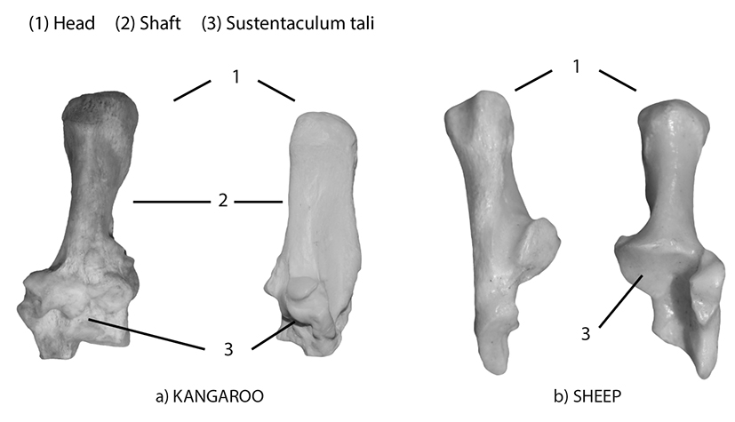
Figure 9.4: Calcanei with diagnostic features labelled in the anterior and medial views; (a) kangaroo, (b) sheep.
Metacarpals and metatarsals (collectively termed metapodials) are the long, tubular bones that connect the wrist or ankle joint with the phalanges (toe bones). Most species have four, but again their number is variable (Figure 9.5). They are larger than, but morphologically similar to phalanges, and have two articular surfaces (proximal and distal). In some species, such as cattle, sheep and horses, the metacarpals and tarsals are reduced in number, reflected in their morphology. In cattle and sheep, the metapodials are reduced to two (the third and fourth metapodials), which fuse together early in life, forming one wide, thickened bone with a characteristic double pulley-shaped distal end (Figure 9.5). In macropods, the metacarpals are similar to humans, but the metatarsals are changed. Macropods have evolved to have one large, central metatarsal, the fourth (IV), with varying numbers of smaller metatarsals. Kangaroos and wallabies have four metatarsals in total, the second and third (II, III) are greatly reduced, long and thin, while the fifth (V) is intermediate in size between the large fifth and smaller second and third (Figure 9.6). The changes in the metatarsals of macropods are an adaptation to their unique pattern of locomotion (i.e. hopping). A selected example of articulated fore and hind limbs have been included at the end of this section.
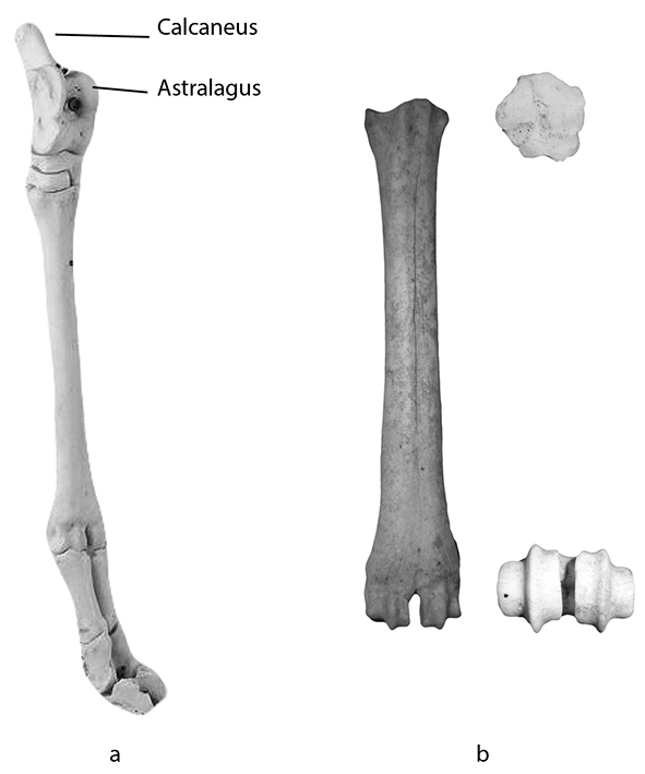
Figure 9.5: (a) Articulated sheep hind foot with calcaneus and astragalus labelled; (b) sheep metatarsal, note the spool-shaped distal end.
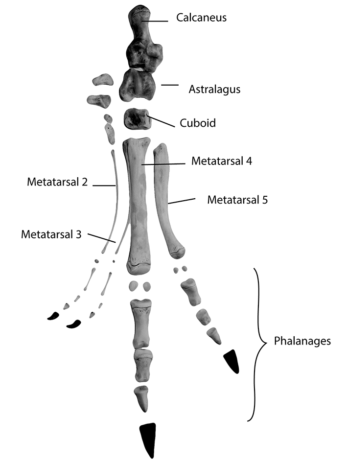
Figure 9.6: Semi-articulated kangaroo foot.
Phalanges (finger and toe bones) follow similar evolutionary patterns as the metapodials. In most species, each digit is comprised of three phalanges (singular, phalanx) (Figure 9.7). These are often referred to as either the first, second or third phalanx, or the proximal, intermediate or distal phalanx. The third, or distal, phalanx of most species is a claw or hoof. It is often very difficult to distinguish between phalanges from the hand and foot, and to avoid certain frustration, this should not be attempted without additional resources.
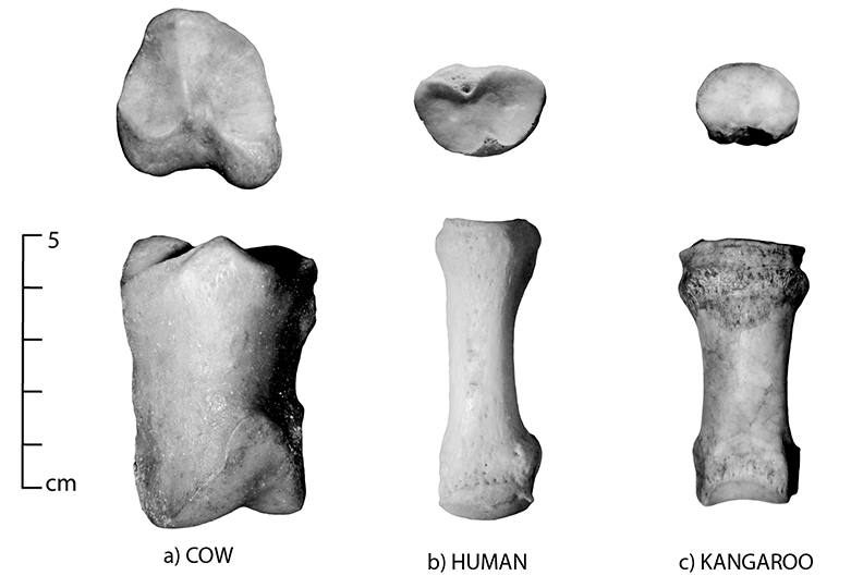
Figure 9.7: Morphological comparison of the first phalanx from (a) cow, (b) human and (c) kangaroo.
Common state in archaeological assemblages
As you can imagine, the morphological variation between the different bones of the extremities plays a role in their preservation. In general, smaller carpals, tarsals and phalanges survive well; however, they are often the subject of recovery bias if an assemblage is not sieved (as smaller bones may be missed when recovered by eye). From the perspective of subsistence, hands and feet are typically less desirable than other, meatier bones such as femurs. This results in variable patterns of consumption, in which their presence or absence can have different interpretations. An absence of extremities, coupled with the presence of long bones, might suggest offsite butchery. In a settlement context, this pattern might also have implications for status. The presence of extremities alone could represent waste or it could represent consumers of lower socio-economic rank if the bones show signs of cooking/butchery.
There are also many uses of metapodial bones in contexts outside of subsistence. In industrial contexts involving hide-working such as those associated with tanning, the large metapodials of many species (cattle, sheep, horse) were left intact with the skin to be used as handles to aid in processing hides. Large metapodials (such as those of sheep, cattle or horses) have had many uses, including roof repair. Astragali were used in gaming in the ancient world (often drilled and filled with iron to weigh them down), and they were even used as paving for floors (along with metapodials). Tools, implements, buttons and all sorts of smaller items have been made from metapodials. Cut marks on most are rare, with the most common being encircling marks on the proximal metacarpals or tarsals, representing skinning. Cut marks may also be found on astragali, resulting from the disarticulation of the foot from the lower leg. Phalanges and smaller carpal or tarsal bones rarely contain marks.
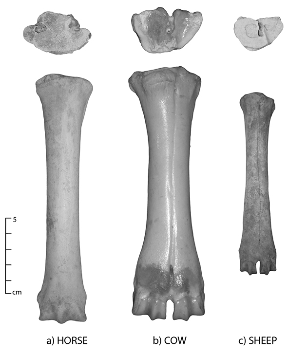
Figure 9.8: Metacarpals; proximal view (top) and anterior view (bottom); (a) horse, (b) cow and (c) sheep. See Figure 2.2 for an image depicting cow metacarpal articulation with the surrounding bones of the fore limb.
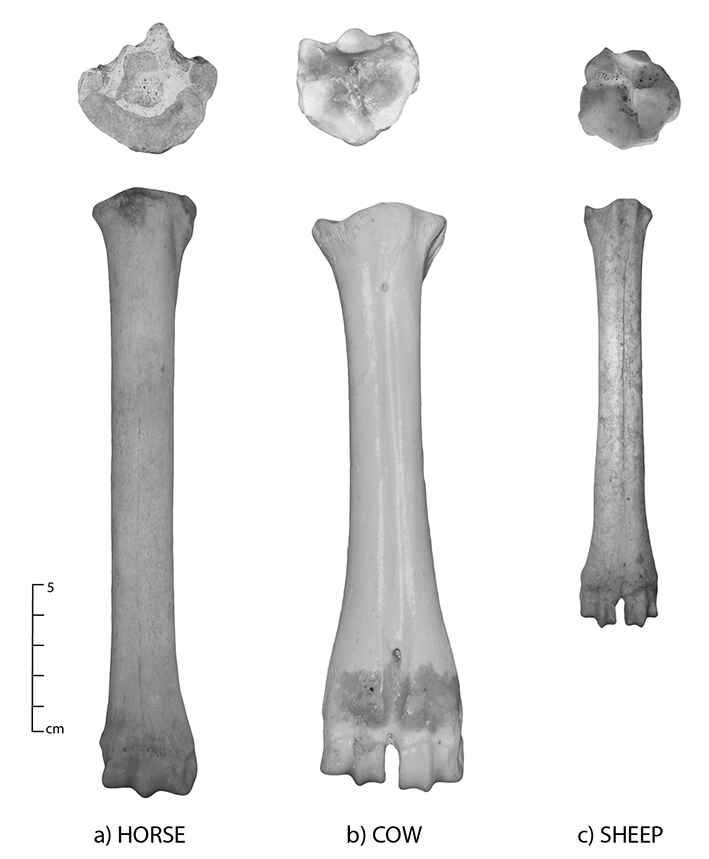
Figure 9.9: Metatarsals; proximal view (top) and anterior view (bottom); (a) horse, (b) cow and (c) sheep. See Figures 9.12–9.14 for images depicting metatarsal articulation with the surrounding bones of the hind limb.
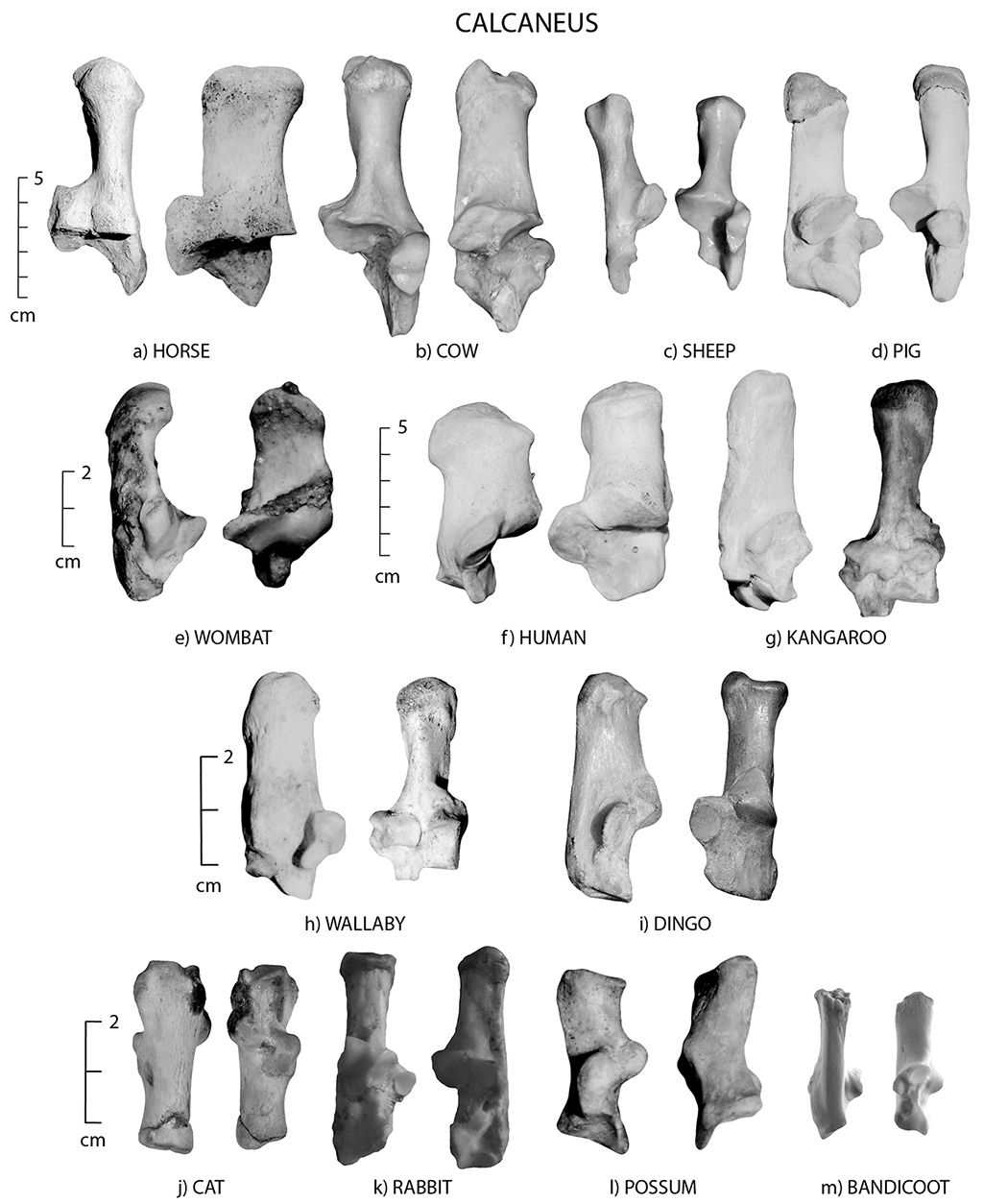
Figure 9.10: Calcanei; medial view (left) and anterior view (right); (a) horse, (b) cow, (c) sheep, (d) pig, (e) wombat, (f) human, (g) kangaroo, (h) wallaby, (i) dingo, (j) cat, (k) rabbit, (l) brushtail possum and (m) bandicoot.
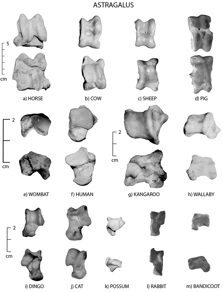
Figure 9.11: Astragali; superior view (top) and anterior view (bottom); (a) horse, (b) cow, (c) sheep, (d) pig, (e) wombat, (f) human, (g) kangaroo, (h) wallaby, (i) dingo, (j) cat, (k) brushtail possum, (l) rabbit (right side) and (m) bandicoot.
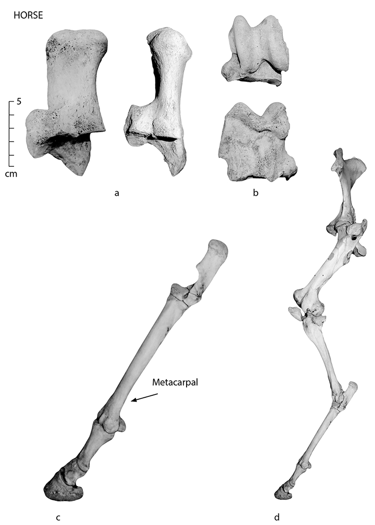
Figure 9.12: Horse (a) calcaneus, (b) astragalus, (c) articulated fore limb and (d) articulated hind limb (see also Figure 7.2). Note: (c) and (d) not to scale.
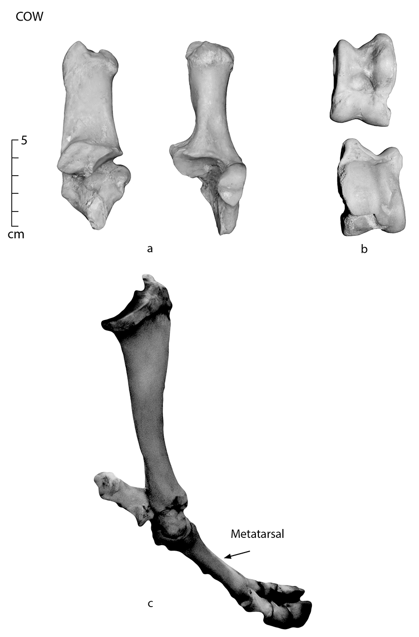
Figure 9.13: Cow (a) calcaneus, (b) astragalus and (c) articulated hind limb.
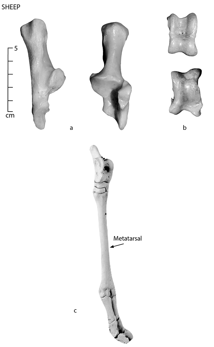
Figure 9.14: Sheep (a) calcaneus, (b) astragalus and (c) articulated hind limb.
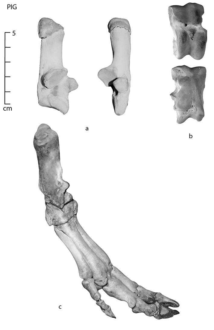
Figure 9.15: Pig (a) calcaneus, (b) astragalus and (c) articulated hind limb.
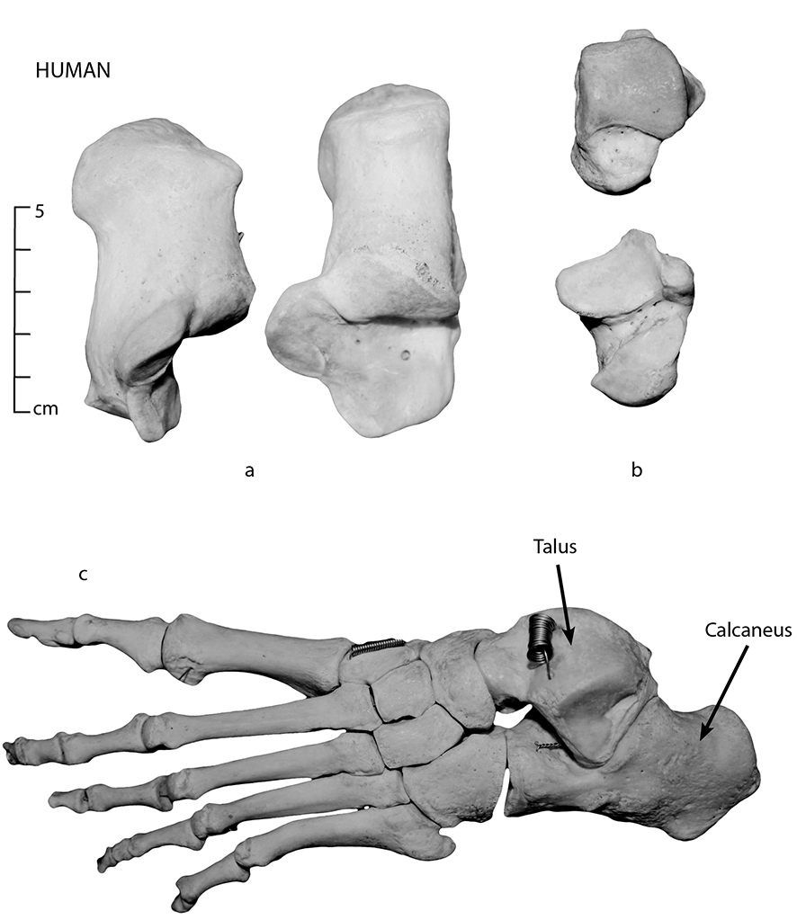
Figure 9.16: Human (a) calcaneus, (b) talus (astragalus) and (c) articulated foot.
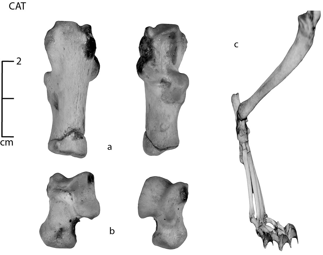
Figure 9.17: Cat (a) calcaneus, (b) astragalus and (c) articulated hind limb.
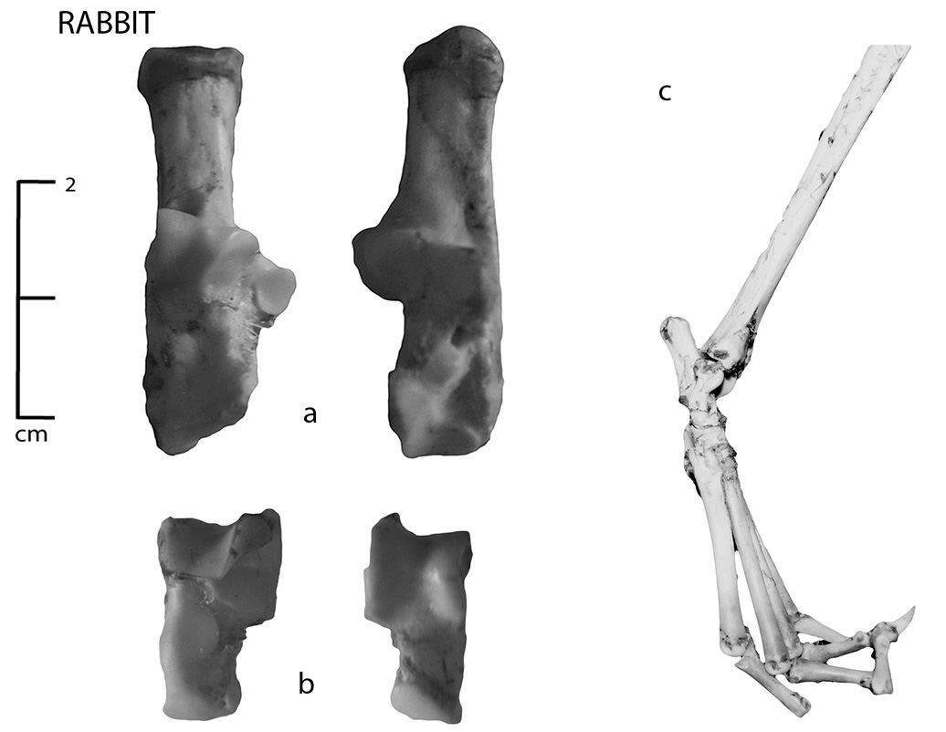
Figure 9.18: Rabbit (a) calcaneus, (b) astragalus and (c) articulated hind limb.
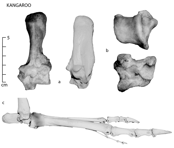
Figure 9.19: Kangaroo (a) calcaneus, (b) astragalus and (c) articulated hind foot (II metatarsal missing from view).
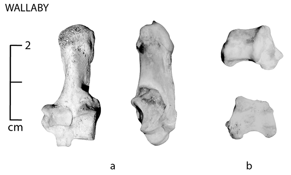
Figure 9.20: Wallaby (a) calcaneus and (b) astragalus (see Figure 9.6 for articulated view).
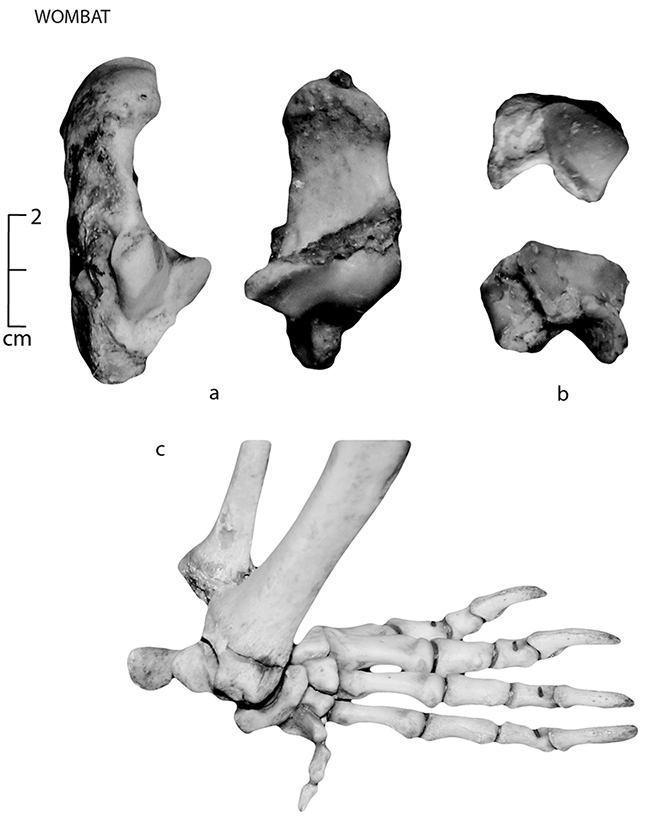
Figure 9.21: Wombat (a) calcaneus, (b) astragalus and (c) articulated hind limb.
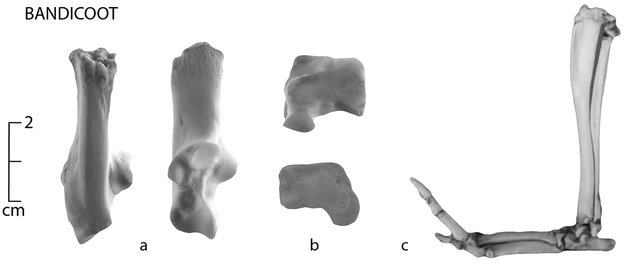
Figure 9.22: Bandicoot (a) calcaneus, (b) astragalus and (c) articulated view.
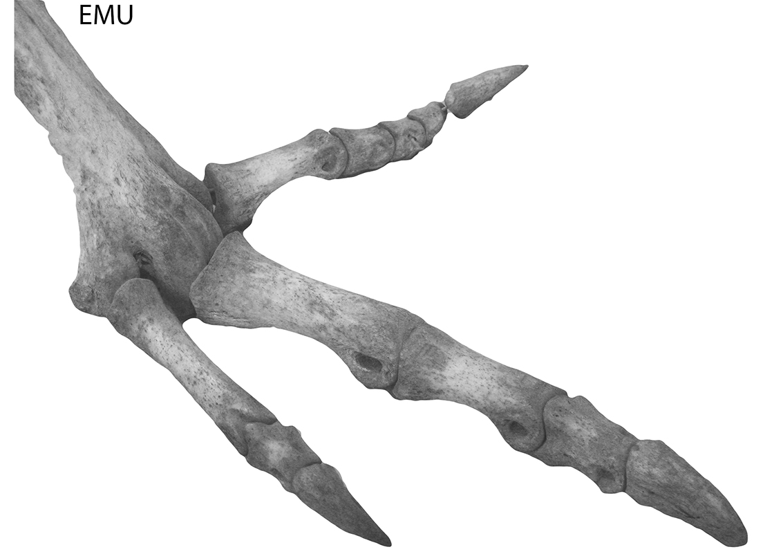
Figure 9.23: Emu articulated limb.