8
Tibia
The tibia, or shin bone, is one of the longest bones in the skeleton of most mammals, and one which is commonly recovered from a variety of faunal assemblages. Together with the smaller fibula, the two bones make up the lower leg, and are analogous to the radius and ulna of the fore limb. In some animal species, the tibia and fibula become fused during adulthood, while in others the fibula has become significantly shortened. The fibula is typically long and thin, and frequently broken in archaeological contexts. While an important bone typically used in the manufacture of bone points, on its own the fibula is difficult to identify without the help of a comparative collection. For this reason, the fibula has been omitted from this manual.
Diagnostic features
Diagnostic features of the tibia are shown in Figure 8.1. The tibia is readily identifiable in bone assemblages due to the unique morphology of its proximal and distal articulations, and the shape of its shaft cross-section. The proximal tibia articulates with the distal femur to form the knee joint. This key weight-bearing joint is reflected in the morphology of the broad proximal tibial surface, specifically in the medial (1) and lateral condyles (2), and the intercondylar eminence (3). The tibial tuberosity (4) rises from the anterior proximal shaft (5) and runs down the length of the bone as a crest, giving the tibia a triangular shape when viewed in cross-section. Distally, the tibia has a swollen appearance for articulation with the either the talus or calcaneus to form the ankle joint of many species. The distal articular surface is trough-like, and is characterised by the medial malleolus (6), a small protrusion or process that in humans forms the ‘bump’ on the inside of the ankle.
Orientation and siding
The tibia is one of the easiest bones to orient and side, due to the distinctive crest, the tibial tuberosity, on its anterior surface. To orient the tibia, place the broad, wide articular surface up or proximal, and the small, trough-like depression down, or distal. Next, place the tibial tuberosity facing away from you – or anterior. When placed in this way (anatomical position), the crest will lean toward the side from which it came.
Species identification
Refer to Figures 8.2–8.7 for species identification using the tibia. While the tibia is one of the easiest bones to identify, it is also one of the harder ones to correctly assign to species as a result of its remarkable uniformity. This said, the two best areas on which to base species identifications are the proximal and distal articular surfaces – with the distal articulation being the ‘easiest’. As with all elements, the morphology of the tibia is a reflection of the way in which an animal moves. This means that the tibias of most quadrupeds will appear similar, as will those of most bipeds. In Australian sites, this translates into kangaroo tibias being frequently mistaken as human. The following list contains some specific inter-species distinctions.
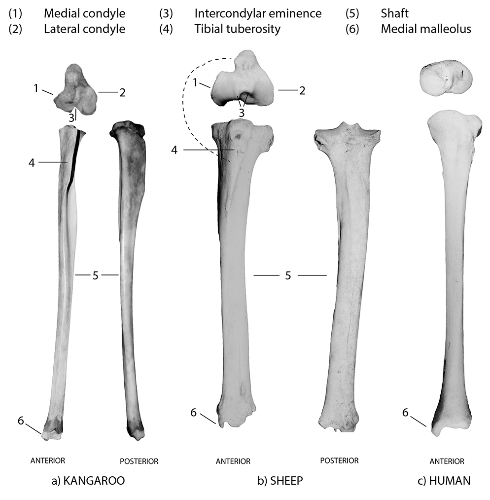
Figure 8.1: Anterior and posterior views of the tibia, labelled: (a) kangaroo, (b) sheep and (c) human.
Distinguishing between humans and animals
- The proximal condyles of the human tibia are rounded in appearance, while they are triangular in most other species (including macropods) (Figure 8.3).
Distal articulation
- The distal articulation of a human tibia is triangular in outline, with very shallow, less pronounced ‘troughs’ for articulation with the ankle (talus). There is a prominent medial malleolus (see Figure 8.3b).
- The distal articulation of a macropod tibia (kangaroos and wallabies) is similar to the human, but has a rectangular outline, rather than a triangular outline (Figures 8.3c and d).
- The distal articulation of cows and sheep has an additional twin-lobed protrusion extending laterally of the main articulation.
Other comparisons
- To distinguish cattle from horses: horses have a groove on the upper-most portion of the tibial tuberosity that produces two distinct ‘swollen’ areas, whereas in cows this area has just one very roughened swollen area (Figure 8.2).
- To distinguish pigs from sheep: pigs have a very short, thick tibia that is robust in appearance and has a pronounced medial malleolus (because pigs have a fibula). Sheep have long, thin tibias with a flared distal end on both sides (Figures 8.4a and b).
- To distinguish wombats from pigs: distally, wombats have a very pronounced medial malleolus that extends beyond the articular surface. Pigs have a pronounced tibial tuberosity, whereas wombats have a sharp crest that extends two-thirds of the way down the shaft, giving the tibia a very flattened, twisted appearance (Figures 8.4b and c).
- To distinguish dingoes from sheep: both have long, thin tibias, but the dingo has a pronounced medial malleolus balanced by a smooth, knob-like lateral end, whereas the distal sheep tibia flares laterally.
- To distinguish rabbits from possums: possums have a proximal surface that is rounded in outline, in contrast to the triangular outline of rabbits. Distally, the rabbit tibia is flared on both sides, whereas the possum tibia ends in a pronounced medial malleolus that has a knob-like appearance.
- The fibula is fused to the tibia in rabbits (Figure 8.5d). It is unfused but greatly reduced in horses, emus and chickens. The fibula is also unfused in kangaroos, wallabies, wombats, humans, pigs, dingoes, cats and possums. The fibula is absent in cows and sheep.
- The tibia of a bird is often referred to as a tibio-tarsus, and is easily recognisable by a distinctive morphology that includes a rounded, spool-shaped distal articulation and a very prominent tibial crest that often rises higher than the proximal articular surface.
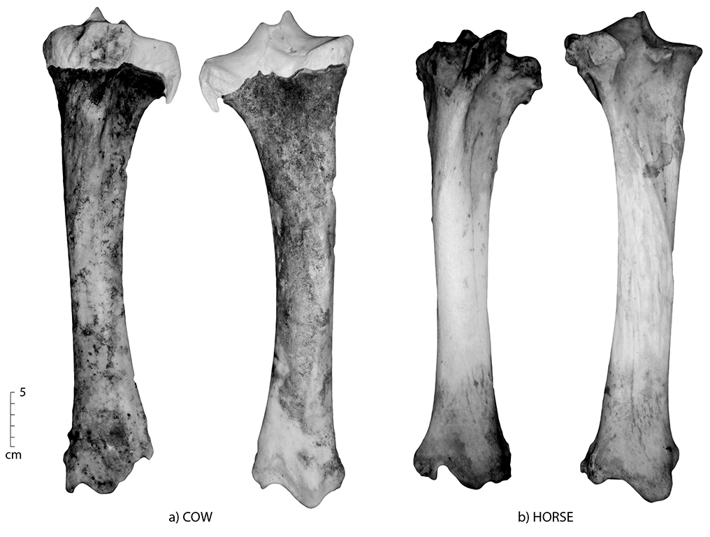
Figure 8.2: Anterior and posterior views of the tibia; (a) horse and (b) cow. Note the groove on the upper part of the tibial tuberosity of the horse that gives two protruding areas.
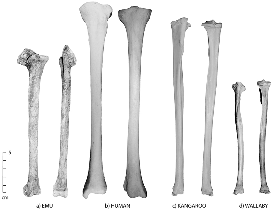
Figure 8.3: Anterior and posterior views of the tibia; (a) emu, (b) human, (c) kangaroo and (d) wallaby. The human tibia is long and slender, with rounded proximal condyles; however, in most other species, the proximal end is triangular.
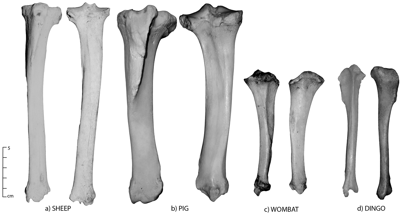
Figure 8.4: Anterior and posterior views of the tibia; (a) sheep, (b) pig, (c) wombat and (d) dingo.
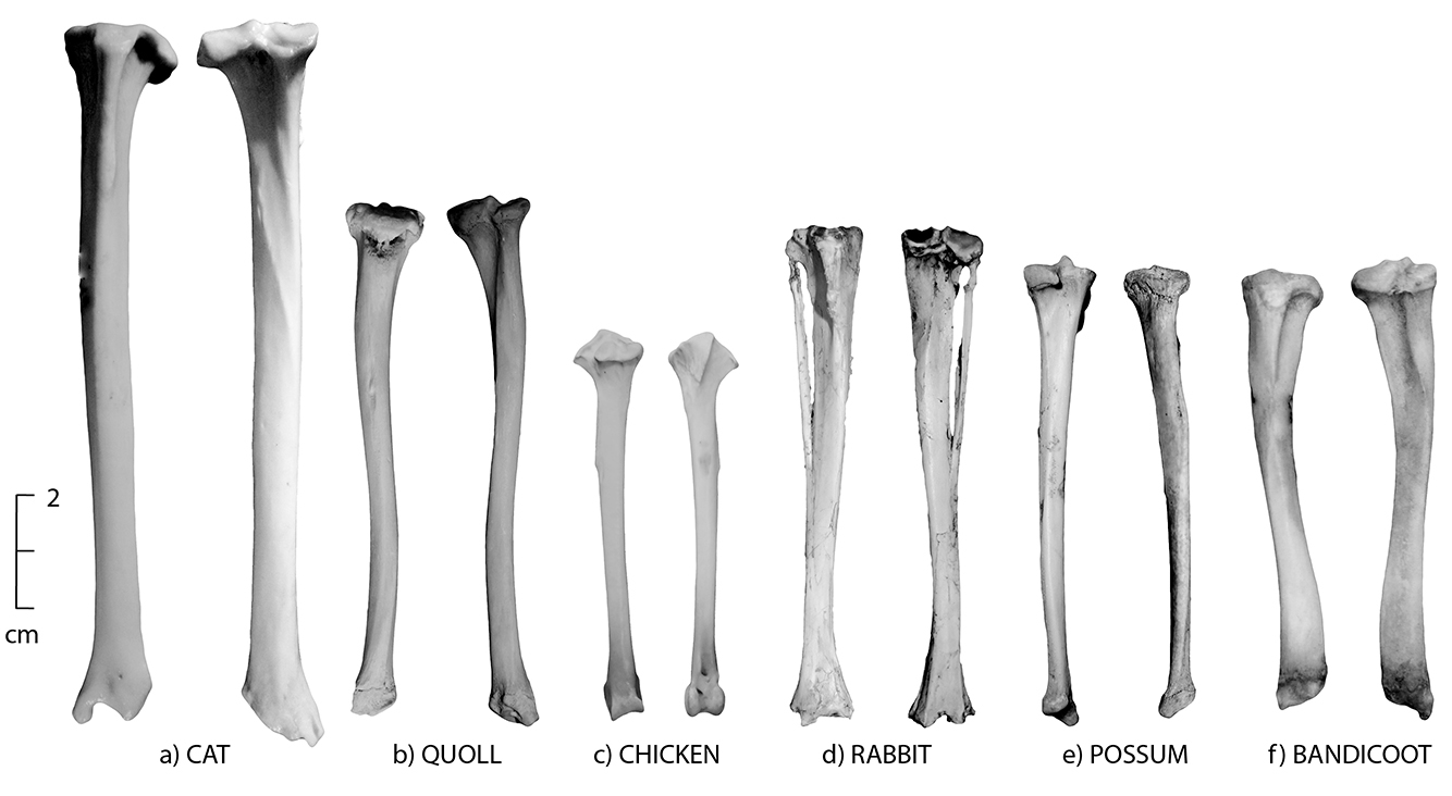
Figure 8.5: Anterior and posterior views of the tibia; (a) cat, (b) quoll, (c) chicken, (d) rabbit, (e) brushtail possum and (f) bandicoot.
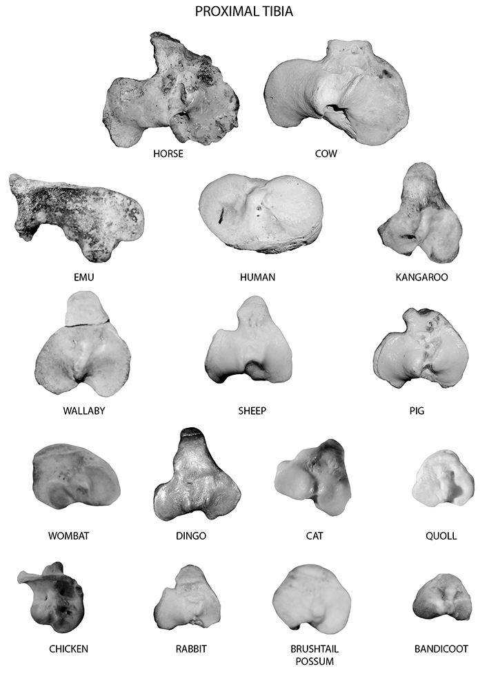
Figure 8.6: Proximal tibia of all species (in order of decreasing size). Smaller species are shown at an exaggerated size to show detail.
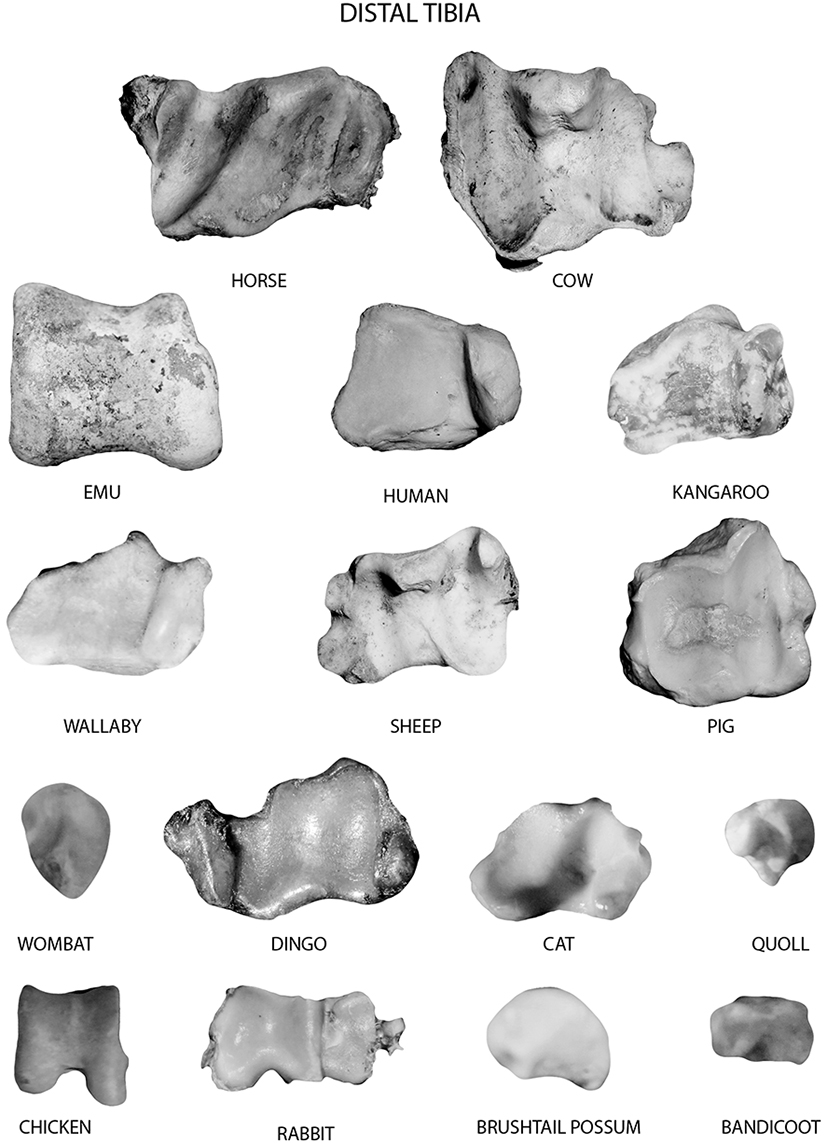
Figure 8.7: Distal tibia of all species (in order of deceasing size). Smaller species are shown at an exaggerated size to show detail.
Common state in archaeological assemblages
The tibia is one of the few bones that may be recovered complete from many archaeological contexts. This may be due to its general robustness or tendency to remain articulated with the ankle and foot bones (especially in macropods and birds). When broken, the fragmentation of the tibia in most species follows a similar pattern. The proximal articulation is often missing, as it is also an epiphyseal cap that in young species may not be fully fused at the time of death. In marsupials, the proximal surface fuses very late in life, and often not at all. The distal articulation of the tibia is also late fusing in marsupials, and often missing unless the bone remained articulated with the foot. In most other species, the tibia may be broken in two around the mid-shaft, frequently resulting in medio-distal fragments that can be identified on the distal morphology and the triangular cross-sectional shape of the shaft.
The relative completeness of the tibia varies between types of sites and assemblages. For example, in naturally accumulated assemblages in which an animal may have died without human agency, the tibia often remains articulated with the foot. This results in the recovery of medio-distal tibia with intact distal articulations. This pattern is common, whether the animal has died from being bogged in soft mud or was killed and scavenged by predators. In prehistoric Indigenous assemblages, the tibia may remain articulated to the foot of many species, especially in large kangaroos and emus, as there is little to no edible meat on this body part. However, tibias in Indigenous assemblages may also be reduced to small fragments as a result of being smashed open for marrow extraction. In historic assemblages, the tibia is frequently cut through the shaft, with traces of the easily identifiable striations of a butcher’s band saw. The placement of the cut is dependent upon the portion of meat desired, and may vary between urban and rural contexts.
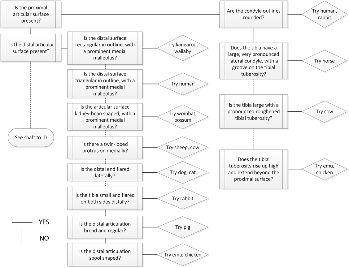 Figure 8.8: Tibia decision process 1. Use this decision process if you have the proximal or distal articular surface of the tibia. |
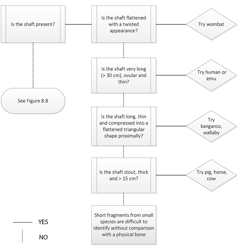 Figure 8.9: Tibia decision process 2. Use this decision process if you have the tibia shaft. |