1
Mandible
The mandible, or jaw bone, is one of the most useful elements in species identification, especially when teeth remain in situ. Just as most major bones in an animal’s body reflect the way in which that individual moves, a species’ dentition and chewing muscles reflect an individual’s diet. As the mandible provides the surface for the attachment of the chewing muscles, as well as the anchor point for the teeth, its morphology can facilitate species identifications at both broad and fine levels. Since there are numerous publications on the identification of teeth (see Hillson 2005), we will devote more attention to the mandible itself, and will conclude with some basic features of dentition that can be used to distinguish between species at a broad level (e.g. herbivore from carnivore, marsupial from placental).
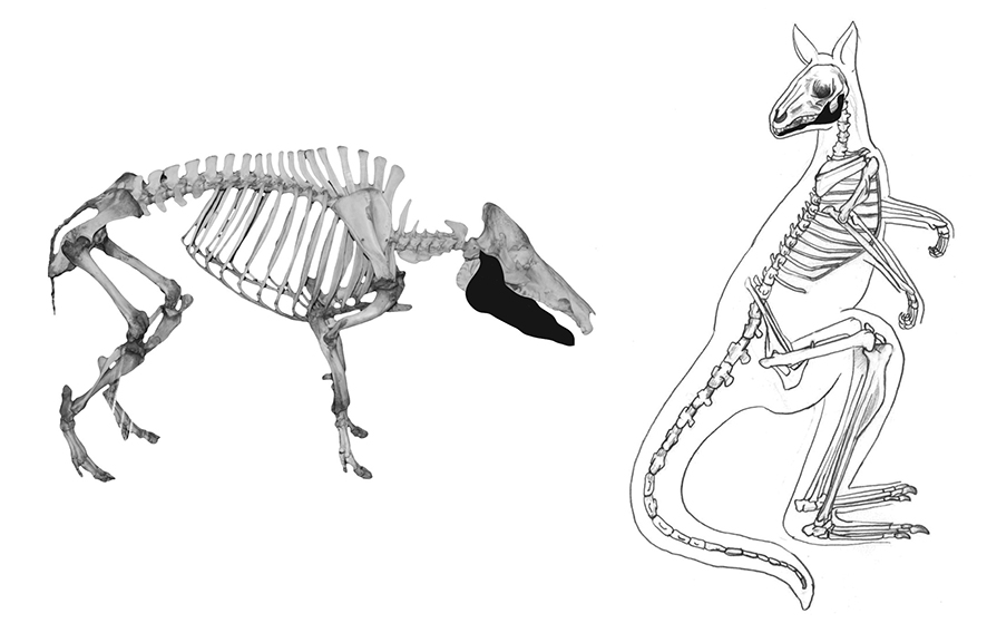
There are morphological differences between the mandibles of marsupial and placental mammals, which means that it is possible to broadly differentiate between these two groups by examining a few specific traits. In particular, the rear portion of the mandible is typically turned inward (inflected jaw angle) in marsupials, as opposed to outward in placental mammals (refer to Figures 0.5 and 1.1). In marsupials, the angular process forms a pronounced shelf that projects medially (lingually) from the posterior mandible (especially notable in wombats). And lastly, the mandibular foramen is an opening that can be viewed from both the buccal and lingual sides in marsupials, as opposed to just the lingual side in placentals (Figure 1.2).
Diagnostic features
Diagnostic features of the mandible are shown in Figure 1.1. The mandible is comprised of two halves (bodies), which join and fuse together anteriorly at the mandibular symphysis (1). Mental foramina (2), small openings in the bone for the insertion of nerves and blood vessels, are present anteriorly on either side of the mandibular symphysis. The mandibular symphysis of many animal species remains cartilaginous throughout life, and therefore in many archaeological assemblages, it becomes ‘unfused’ over time, resulting in just one side or body being recovered. The largest portion of the mandibular body is the ascending ramus (3), a broad, ‘flattened’ area of the distal mandible. Typically, animals engaged in heavy chewing have a larger ramus. Extending upward from the ascending ramus is a smooth, rounded (either concave or convex) articular surface, called the mandibular condyle (4), which forms the temporomandibular joint with the cranium. The coronoid process (5) also extends upward from the ramus, opposing the mandibular condyle, and serves as the point of attachment for the temporalis muscle. The coronoid process and mandibular condyle are separated by the mandibular notch (6). The masseteric fossa (7) on the buccal (cheek) side of the ramus, is an indentation in which sits the masseter muscle. On the lower distal portion of the mandible, the angular process (8) is an outward projection found in many species, including some marsupials and carnivores, and serves as another attachment point for the masseter muscle. Also on the buccal side, the morphology of the mandibular shelf (9) and location of the mandibular foramen (10) can be used to distinguish between species. The length and location of the diastema (11), a space commonly found before the cheek tooth row on most mammals, is also diagnostic of species.
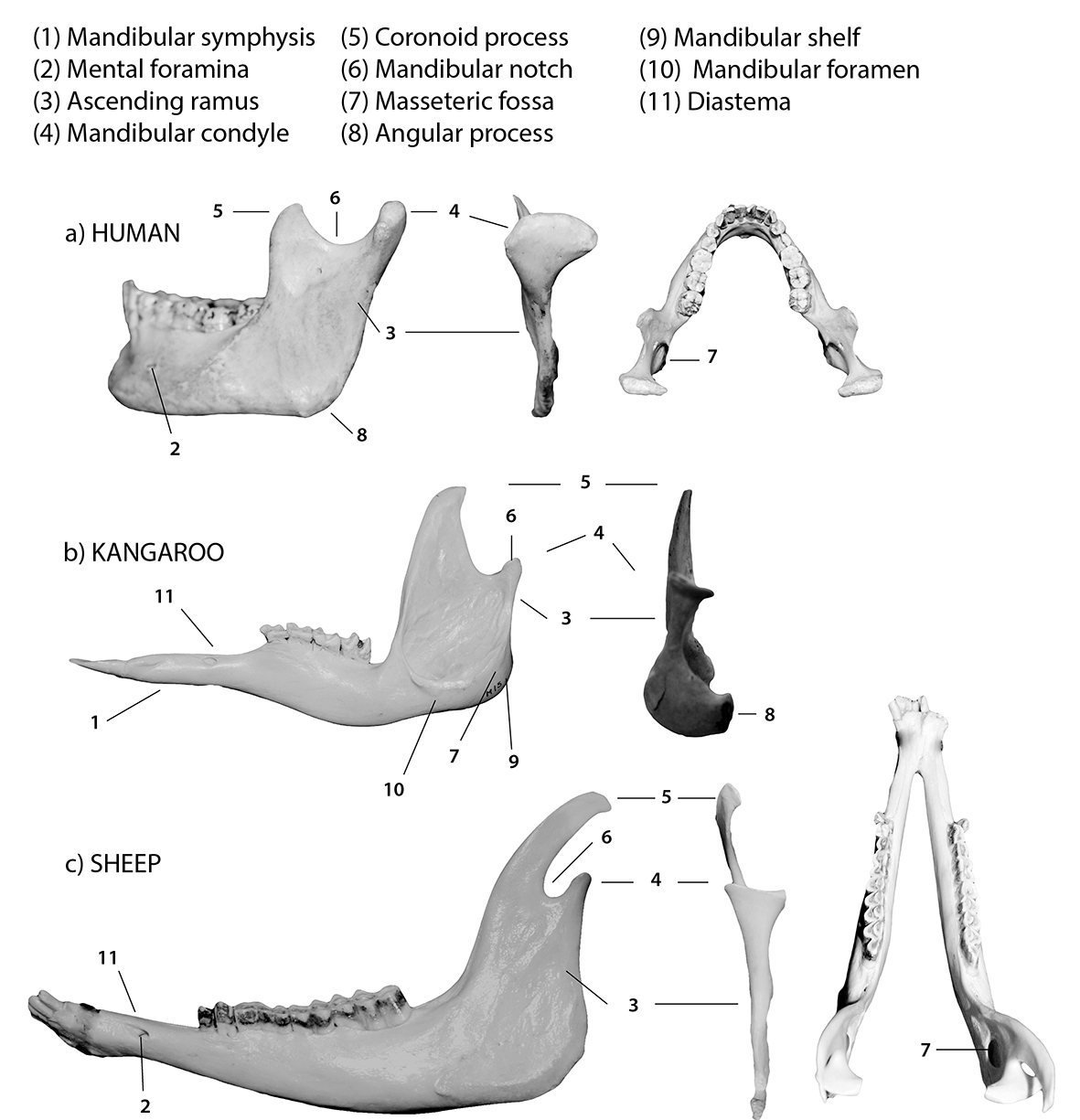
Figure 1.1: Mandibles labelled with diagnostic features; (a) human, (b) kangaroo and (c) sheep; the buccal, posterior and superior (occlusal) views are shown for the human and sheep (L to R).
Orientation and siding
To orient one side (body) of the mandible, the roughened area at the mandibular symphysis indicates the anterior portion of the bone, with the alveoli or in situ teeth facing up (cranially). Any mental foramina are generally anterior and lateral (buccal). The ascending ramus, coronoid process and mandibular condyle are posterior (toward the back).
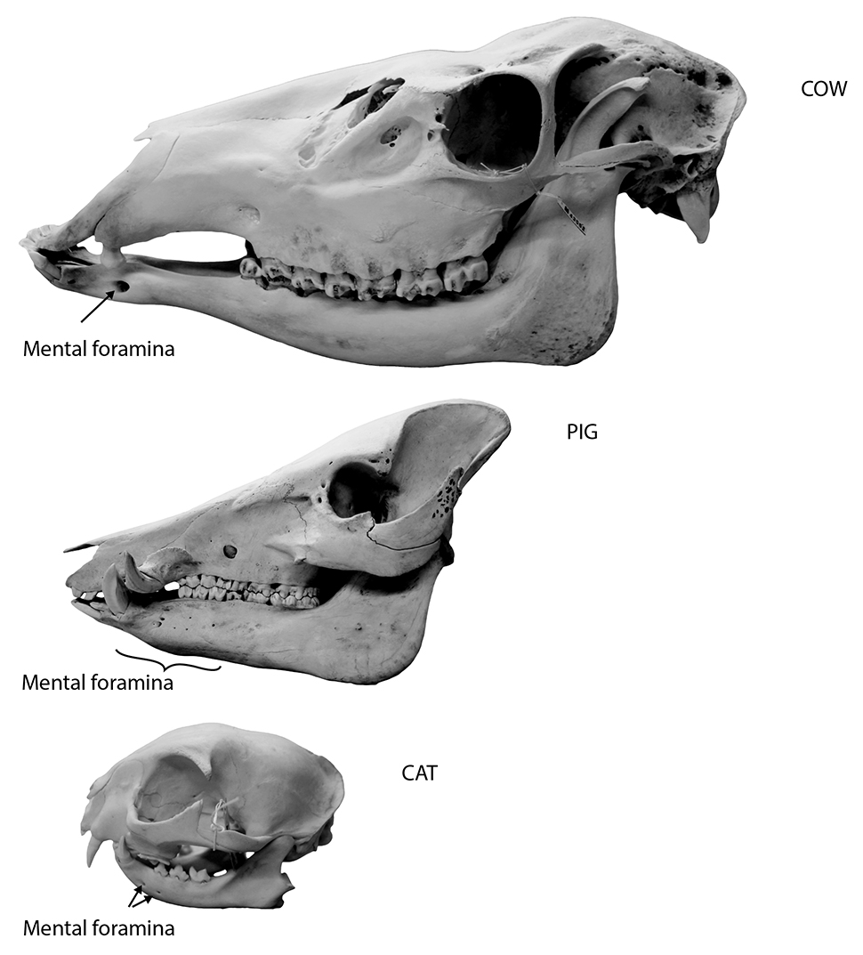
Figure 1.2: Comparison of the mental foramina of a cow, pig and cat.
Species identification
Refer to Figures 1.3–1.8 for species identification using the mandible. If a mandible has teeth in situ, identification to species is much easier. In the absence of dentition, and assuming the mandible is fragmented, the following traits can be used for identification.
Distinguishing between humans and animals
- Humans have a chin; the mandibular symphysis remains cartilaginous, and therefore unfused in most animals, but is replaced with bone (and therefore fused) early on in humans.
- Humans have a U-shaped dental arcade (when viewed from above), visible in intact mandibles (Figure 1.1a).
The size, shape and angle of the ascending ramus
- Dingoes and cats both have a distinct angular process posteriorly (toward the back).
- Marsupials (kangaroos, wallabies, wombats, possums) all have a distinct mandibular shelf lingually (tongue side).
- Pigs and horses have a curved distal ramus that is roughed in appearance, with strong ridge marks.
Morphology of the mandibular condyles
- Dingoes, cats, horses, pigs and humans all have rounded condyles with a ‘rolled’ or convex appearance.
- Wombats have rounded condyles that slope inward (lingually).
- Rabbits have small, ball-like condyles.
- Possums have rounded condyles that are oblong bucco-lingually (cheek to tongue side).
- Cattle, sheep, kangaroos and wallabies all have flattened, saddle-shaped (concave) condyles.
Morphology of the coronoid process (when viewed from the side)
- Dingoes and cats both have a substantial, wide coronoid process.
- Pigs have a low, pointed process that curves posteriorly.
- Humans have a process that is intermediate in height and almost triangular in shape with a very wide mandibular notch.
- Horses have a long, high, almost rectangular-shaped process with a rounded proximal edge that is angled posteriorly (but not curved).
- Cattle also have a long, high process, but one that curves proximally posteriorly.
- Sheep have a prominent, arched process that curves posteriorly.
- Kangaroos and wallabies have a process that is intermediate in height, and curved posteriorly with a shark fin-like appearance.
- Wombats have a process that is tall, curved slightly posteriorly, and culminates in a sharp point.
- Rabbits and possums are similar in size, but the rabbit does not have a process, while the possum has a triangular process with a straight posterior border.
Location of mental foramina
- Most species have one foramen on the buccal anterior surface of each mandibular body.
- Cats and dingoes have two foramina.
- Pigs and rabbits have numerous foramina (Figure 1.2).
Morphology of the mandibular notch
- Humans have a notch that is very wide and open, whereas in most other species, the notch is pronounced (compare Figures 1.3b and c).
- Rabbits have no notch.
- Possums have an L-shaped notch.
- Kangaroos have a notch that appears like a curved L shape that is slightly slanted anteriorly.
- Wallabies have a semilunar shaped notch.
- Wombats have a notch that is a rounded V shape.
- Pigs, dingoes and cats have a very shallow notch.
- Cattle, horses and sheep all have a deep, rounded notch.
The pattern of alveoli (tooth sockets) can be used for species identification (see Hillson 1992), especially the position and length of the diastema (space) between the canines and premolars (or between the incisors and premolars).
The masseteric fossa is very deep and pouch-like in large marsupials (wombats, kangaroos, wallabies).
Distinguishing between kangaroos and wallabies
- In complete mandibular bodies, the curvature of the diastema (the space between the incisors and premolars) can be used to differentiate between macropods.
- Kangaroos have a diastema that curves upward, toward the tooth row, giving the teeth the appearance of sitting higher than the anterior portion of the mandible (Figure 1.4b).
- Wallabies have little to no curvature of the diastema, giving the tooth row the appearance of sitting on the same level (Figure 1.4c).
Common state in archaeological assemblages
The mandible of most animal species is commonly recovered unfused in most archaeological assemblages. Therefore, the morphology of the ascending ramus, mandibular condyles and, if intact, the coronoid process, can be used for species identification. If the mandible is fragmentary, it may be possible to use the location, number and size of the mental foramina and the mandibular symphysis for identification. The mandible is sometimes confused with an innominate when fragmented, as both elements are irregularly shaped. Cut marks are often found on the mandibular condyle, resulting from disarticulation from the skull, and may also be found on the buccal surface resulting from tongue removal.
Teeth tend to preserve well in most environments, with the teeth of smaller individuals often faring the best. Large premolars and molars from horses, cattle and even sheep or goats and marsupials commonly break, with the enamel often shearing off. When this occurs, it may be difficult to distinguish between horse and cattle, and even between cattle and sheep. Isolated teeth belonging to either kangaroos or wallabies are also frequently difficult to distinguish, as the shape of the mandible is the best indicator to distinguish between several species of macropods (Figures 1.4b and c).
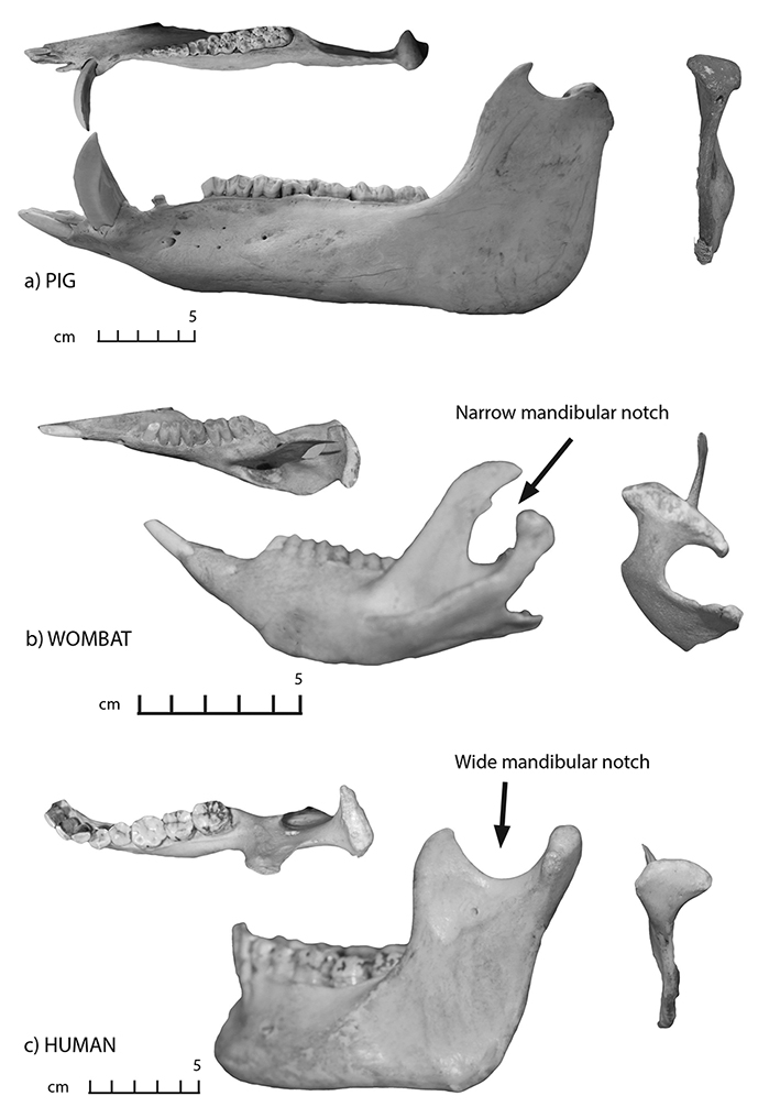
Figure 1.3: Mandibles in buccal and posterior views; (a) pig, (b) wombat and (c) human.
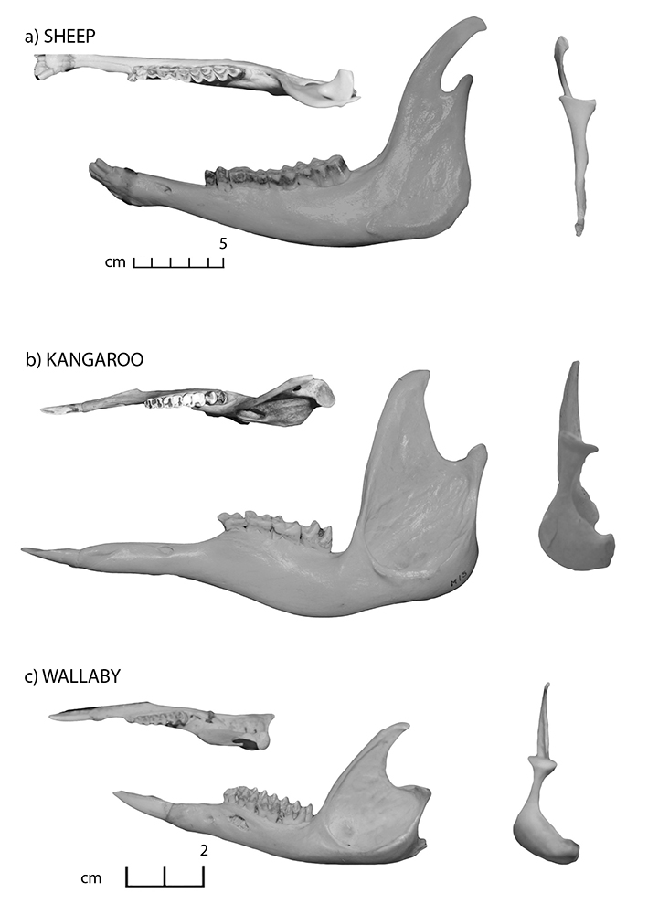
Figure 1.4: Mandibles in buccal and posterior views; (a) sheep, (b) kangaroo and (c) wallaby.
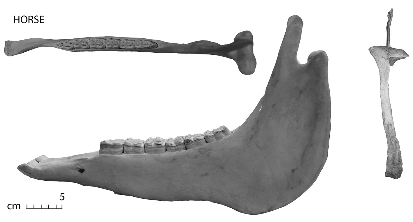
Figure 1.5: Horse mandible in buccal and posterior view.
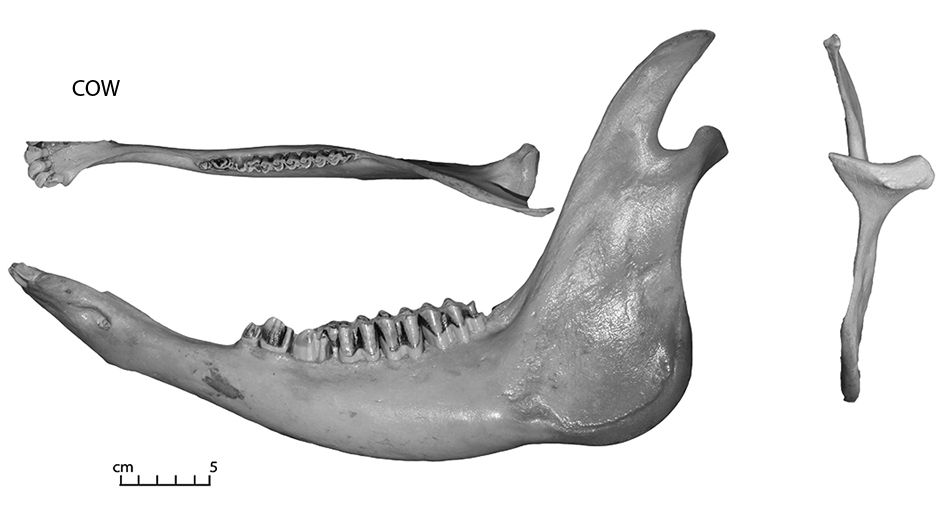
Figure 1.6: Cow mandible in buccal and posterior view.
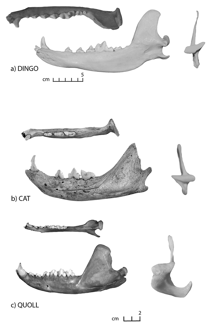
Figure 1.7: Mandibles in buccal and posterior views; (a) dingo, (b) cat and (c) quoll (buccal image right side of animal).
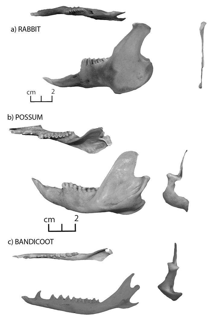
Figure 1.8: Mandibles in buccal and posterior views; (a) rabbit, (b) brushtail possum and (c) bandicoot.
Dentition
The teeth of all species have distinct morphological traits that are extremely useful in identification. All mammals have the same basic types of teeth – incisors, canines, premolars and molars, the shape of which varies between species largely due to dietary differences. All animals (including humans) can be classified according to one of several broad dietary preferences: herbivore, folivore, omnivore, carnivore and insectivore. Horses, cattle, sheep, rabbits, kangaroos, wallabies and wombats are grazing herbivores, subsisting largely on grasses, while goats are browsing herbivores, eating a variety of grasses, leaves, bark and shrubs. Possums are largely folivores, eating mainly leaves, while dingoes, quolls and cats are carnivores, subsisting largely on meat. Humans, pigs and bandicoots are omnivores – eating most things that come their way. Dietary differences are generally best reflected in the premolars and molars (cheek teeth), as these teeth tend to do the majority of the grinding, tearing and shearing needed to process different food sources. In the images found on the following pages, the occlusal (biting) surface of the teeth is illustrated, as this surface contains the clearest morphological differences useful in distinguishing between species. For detailed discussions and information on the dentition of various species, see Hillson (2005, 1992).
Along with morphological differences, every species has a unique dental formula – that is, a specific number of each of the major types of teeth (incisors, canines, premolars and molars) – that is useful in identification (see Figure 1.9). Dental formulas are expressed by taking into consideration one half of the entire mouth and counting the number of each type of tooth. For example, the original mammal dentition formula has the same number of teeth on the top and bottom, and is written 3.1.4.3 / 3.1.4.3. Starting from the midline, this translates into three incisors, one canine, four premolars and three molars on each half of the upper and lower jaws. An easier example to visualise may be the human dental pattern – 2.1.2.3 / 2.1.2.3. This translates into two incisors, one canine, two premolars and three molars in each quadrant of the human mouth. However, some species have different numbers of upper and lower teeth. For example, both bovids (sheep, goat, cattle) and cervids (deer, antelope) do not have upper incisors; bovids also lack upper canines. The following are the dental formulas for the species covered by this manual, followed by some general dental morphological differences between species that may be useful as a starting point in identification. Please note, however, that tooth morphology rapidly changes over an individual’s lifetime due to age or wear, and whether the tooth in question is deciduous (milk tooth) or permanent. Given this large morphological variability in teeth, comparative reference materials should be consulted for accurate identification.
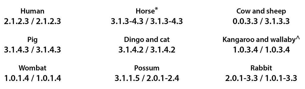
Figure 1.9: Dental formulas (incisors, canines, premolars and molars) for some of the species in this manual. *Some horses have vestigial first premolars called wolf teeth. ^The third premolar is the only deciduous tooth in marsupials, leading to what is often termed ‘molar progression’, whereby it is eventually lost later in life. In many marsupials, molar progression makes distinguishing between premolars and molars a less than clear-cut task. Incidentally, the presence or absence of the third premolar may be used in age estimates.
Marsupials differ from placentals in their dentition. In general, marsupials have more teeth than placental mammals, often having three premolars (PM) and four molars (M) on each side in both the upper and lower jaw (PM 3/3; M 4/4). In placental mammals, the general dental formula is four premolars and three molars on either side in both the upper and lower jaw (PM 4/4; M 3/3).
Species identification
Refer to Figures 1.3–1.8 for species identification using dentition.
| Horse | Cheek teeth (molars and premolars) are rectangular in outline (as opposed to tapered), blocky, tall, and all similar to one another in form; upper and lower incisors are conical in form (Figure 1.5). |
| Cow, sheep and goat | Cheek teeth are high-crowned, with deep infoldings (crescent moon appearance); lower incisors and canines are spatulate in form (Figure 1.6). |
| Kangaroo and wallaby | Cheek teeth are characterised by an H pattern on the occlusal surface; lower incisors are large, pointed, and projecting with small, spatulate upper incisors (Figure 1.4). |
| Wombat | Cheek teeth are blocky with an M-shaped outline; incisors are large and projecting (almost tusk-like). Their rootless teeth grow continuously (Figure 1.3b). |
| Cat, dingo and quoll | Cheek teeth are sharp, and blade-like with several points; incisors are spatulate and pointed; canines are sharp and pointed (Figure 1.7).
|
| Human and pig | Cheek teeth are rounded and broad in profile with several cusps; incisors are spatulate, while canines are pointed (male pig canines are tusks). Pig and human teeth are commonly mistaken for each other, but can be easily differentiated by their cusp patterns (Figures 1.3a and c). |
| Rabbit | Cheek teeth are ridged and blocky, all of similar size; incisors are large and projecting (Figure 1.8a). |
| Possum | Cheek teeth are pointed, comprised of two parallel crests, separated by a trough; lower incisors are large and projecting (growing throughout life), while upper incisors are small and spatulate (Figure 1.8b). |
| Bandicoot | Cheek teeth are distinctly ‘pointed’ on the buccal side; premolars are sharp and pointed (Figure 1.8c). |
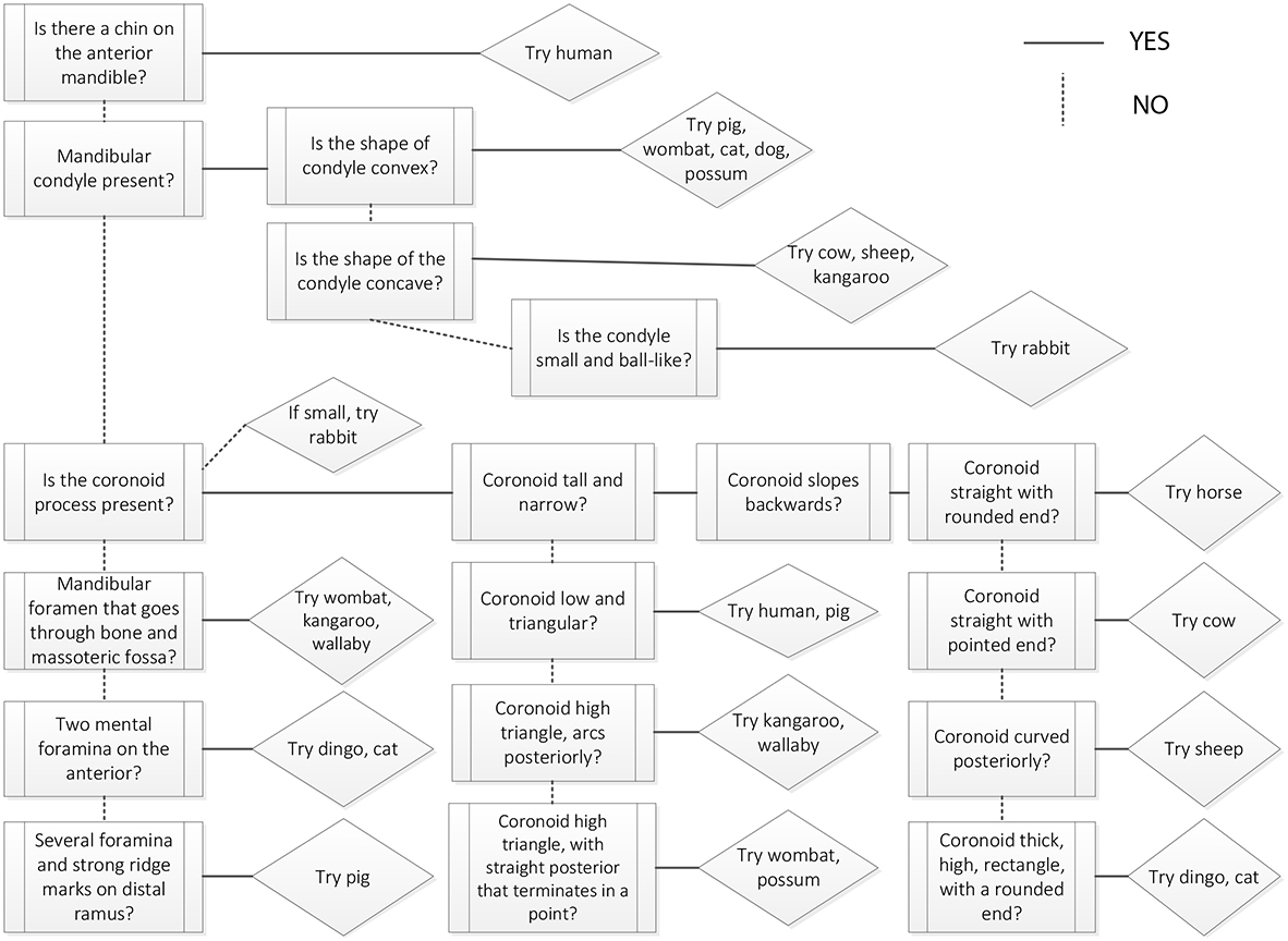 Figure 1.10: Mandible decision process. |