6
Pelvis
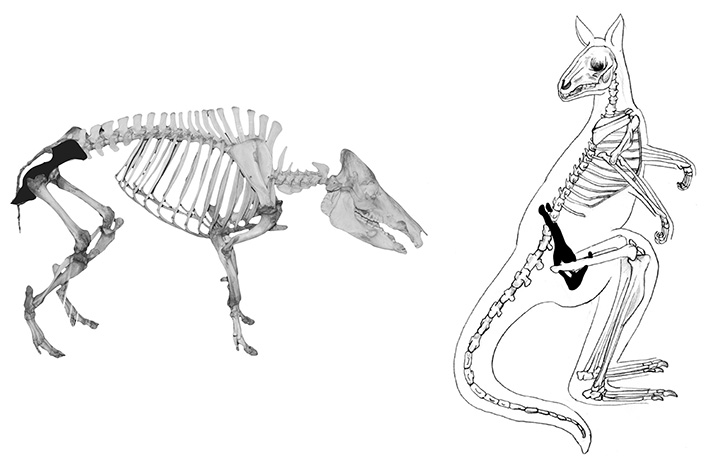
The pelvis is made up of three bones, a left and right os coxa (1) (more commonly referred to as the innominate) and the sacrum, which joins the two sides (see Figure 6.1). In most species, these three bones fuse together to form the hip or pelvic girdle, but are recovered disarticulated in the majority of archaeological contexts. Since the innominate is the most readily identifiable part of the pelvis, this bone will be the focus of discussion.
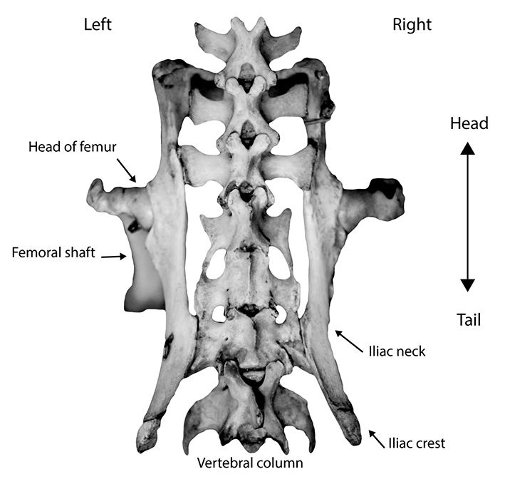
Figure 6.1: Articulated pelvis and sacrum of the quoll.
Diagnostic features
Diagnostic features of the pelvis are shown in Figure 6.2. Each innominate (or half) is comprised of three fused bones – the ilium (2), ischium (3) and pubis (4). One of the most diagnostic parts of the innominate is the acetabulum (5), or hip socket, and the acetabular notch (6), into which fits the head of the femur to form the hip joint. Due to its robust morphology, the acetabulum is nearly always intact if extant in archaeological assemblages. Less frequently, the entire innominate (left or right half) remains intact, in which case the shape of the obturator foramen (7), the sciatic notch (8), the pubic symphysis (9), the height of the iliac neck (10), the shape of the iliac crest (11) and the depth of the supra-acetabular fossa (12) may also be used in species identification.
In addition to definitive species identification, the pelvis is also a key bone in the determination of sex and age, especially in humans. The biological ability to bear offspring has a direct effect on the morphology of the pelvis of nearly all mammalian adult females. Morphological differences are best identified with intact pelvic girdles, although it is often possible to sex even fragmentary elements. In humans, the size and shape of the ilium, opening of the pelvis girdle, and morphology of the pubic symphysis and sciatic notch are all indicators of sex. Age and number of births may also be determined by the morphology of the pubic symphysis. References that contain further information on ageing and sexing the pelvis can be found at the end of this manual.
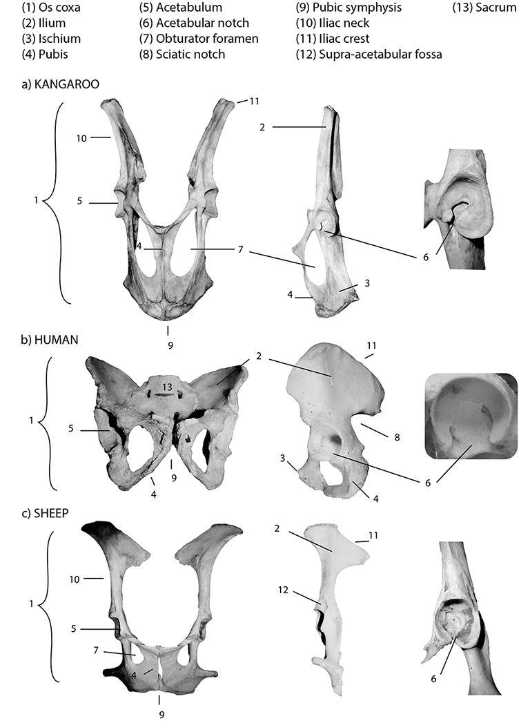
Figure 6.2: Pelves with diagnostic features labelled; (a) kangaroo, (b) human and (c) sheep. Details from left to right: articulated pelvis, os coxa and acetabulum. Note that the human pelvis has been rotated slightly and shown articulated with the sacrum.
Orientation and siding
To side and orient a complete pelvis, hold the pelvis with the fused pubic symphysis facing away from you (anterior), and the sacrum facing you (posterior). Orient the pelvis so the acetabula are facing laterally, and the ilia are pointing up (cranially). In this position, a left innominate will be on your left, etc. If just one innominate is extant, orient it by placing the ilium/iliac crest cranially, and the pubic symphysis/obturator foramen caudally. The acetabulum and ischium should be lateral, and the pubic symphysis medial.
Species identification
Refer to Figures 6.3–6.11 for species identification using the pelvis. Several morphological characteristics of the innominate facilitate species identification, however the most robust of these is the acetabulum. For this reason, the decision process at the end of this chapter is based on the morphology of this element. As with all bones, the size and shape of the pelvis is a reflection of the way in which an animal moves and the degree to which their young are born altricially (incapable of moving around) or precocially (quickly mobile). Altricial human babies are born with large heads relative to the rest of their bodies, due to larger brain size. The need to fit a grapefruit-sized object through a narrow opening, coupled with bipedal locomotion (walking on two legs), is a governing factor in the relatively low, rounded shape of the human pelvis (which translates into a wide ilium in the innominate). Conversely, most animals in this manual are precocial quadrupeds that bear young with relatively short maturation periods and smaller heads and brains. These traits are exemplified by tall, narrow, and often distally flared iliac blades, among other characteristics. Marsupials, who bear undeveloped pouch young, have epipubic bones (see Figure 0.6 in ‘Bone identification 101’). The depth of the acetabulum is also an indicator of locomotion and habitat.
As the size and general shape of the pelvis varies considerably between species due to all of these factors (and more), several different views of the pelvis are depicted here. The acetabulum and iliac neck and crest is shown for every species, and also included are views of several articulated pelves to illustrate variability in size and shape across a variety of species (Figure 6.3).
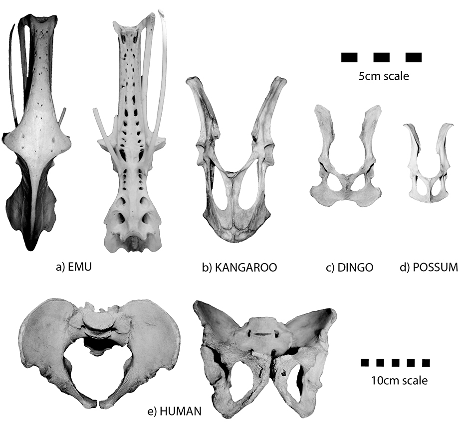
Figure 6.3: A size and shape comparison of several different articulated pelves; (a) emu, (b) kangaroo, (c) dingo, (d) possum and (e) human.
The following are some specific morphological distinctions (refer to Figures 6.4–6.11).
Acetabular notch
- The acetabular notch is open for humans (see Figure 6.6a), horses (Figure 6.4a), wombats, dingoes, cats and possums.
- The acetabular notch is almost closed for cattle (see Figure 6.4b), sheep, pigs, kangaroos, wallabies and rabbits (Figure 6.5).
- The acetabular notch is widely open, giving the acetabulum a U-shaped outline in humans (see Figure 6.6a), dingoes (Figure 6.6c) and cats.
- The acetabulum lacks a ‘notch’, and is smooth and round, resembling a ring in emus (see Figure 6.4c) and chickens.
- Cattle and horses have a large acetabulum (> 50 mm in diameter), the acetabulum of pigs, humans and kangaroos is medium (30–50 mm), the acetabulum of wallabies, wombats, sheep and dingoes are small (15–30 mm), and those of cat, rabbit and possum are all very small (generally < 15 mm).
- Pigs have a very raised acetabular rim when viewed from the side, with a very deep fossa.
Supra-acetabular fossa
- Cattle and sheep have a deep, slit-like supra-acetabular fossa, however in other species the supra-acetabular fossa is very shallow and more like a depression than a fossa.
Pelvic neck and blade
- The neck of the ilium is broad relative to its length, terminating cranially in a rounded, paddle-shaped iliac crest in dingoes, cats and rabbits (see Figures 6.8c, d and f).
- The neck of the ilium is long and thin, terminating in a flared, almost triangular iliac crest: horse, cow, sheep and pig (see Figures 6.9 and 6.10b).
- The neck of the ilium is long and tapers gradually toward a narrower iliac ‘crest’ in kangaroos, wallabies and possums (see Figures 6.8a, b and g).
- In wombats, the neck of the ilium is long, slender and terminates in a sickle-shaped iliac blade that curves laterally (Figure 6.10c).
- The innominate is long and irregularly shaped, curved cranially and characterised by many holes ventrally (resembling a ‘Swiss cheese’ appearance), with a rounded acetabulum in the centre in emus and chickens (Figure 6.11).
Differentiating between marsupial and placental mammals
- If a complete innominate is present, marsupials are relatively easy to distinguish from placentals by the long, thin, cranially tapering ilia.
Common state in archaeological assemblages
Innominates are frequently recovered from archaeological assemblages, but generally in a fragmentary state. When incomplete, parts of the pelvis are commonly mistaken for other elements. For example, the ischium of some species is often confused with the mandibular ramus, while the neck and iliac crest can be confused with the scapula. In these cases, use of a comparative collection is the best way to make a positive identification. The pelvis is almost always broken as a result of either natural (scavenging, post-depositional disturbance) or anthropogenic factors (for example, butchery). In naturally accumulated faunal assemblages, scavenging species tend to disarticulate and break the pelvis early on when accessing a carcass. Carnivore damage often results in a broken ilium, resulting from gnawing and accessing the internal organs. The ischium and pubis may also be broken during these processes. The acetabulum generally survives due to its general robustness. In historic anthropogenic (human-accumulated) faunal assemblages, portions of the innominate are often recovered as they form part of common leg meat cuts (along with the femur and tibia). In prehistoric assemblages, the bone is also frequently extant. Cut marks may be found in or around the acetabulum as a result of the disarticulation of the femur, as well as on the iliac blade if skinning has occurred. Band saw cuts are also commonly found on either side of the acetabulum.
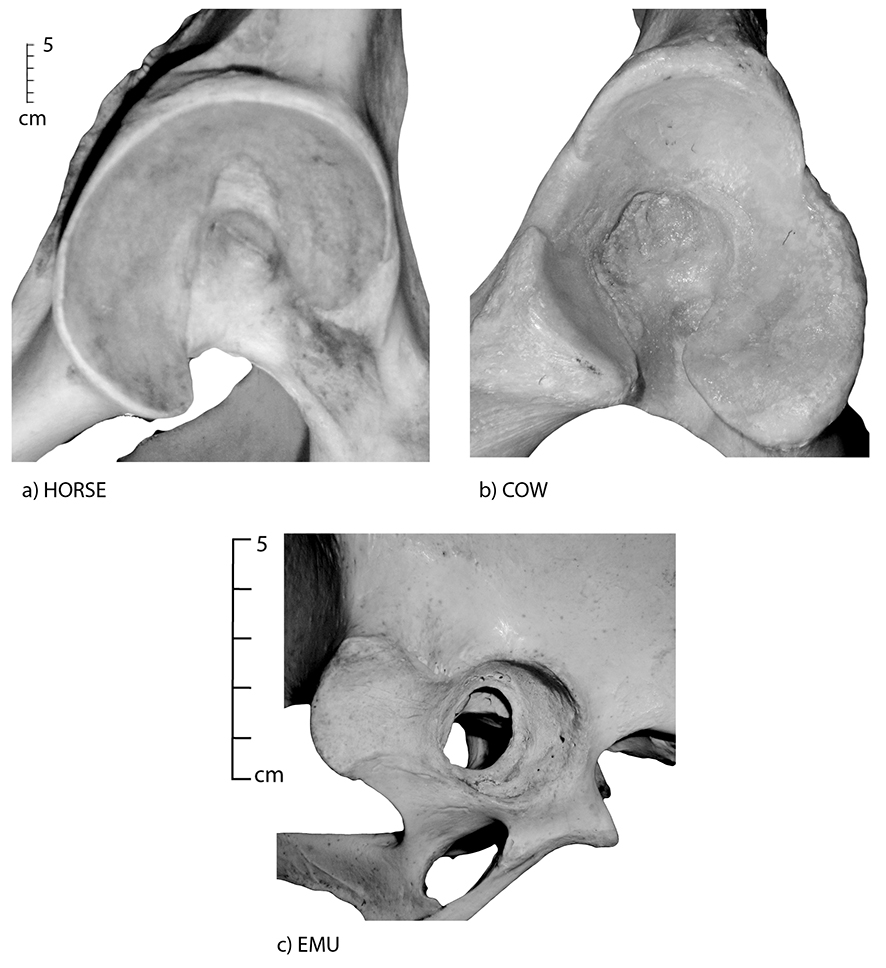
Figure 6.4: A detail image of an acetabulum; (a) horse, (b) cow and (c) emu; illustrating an open acetabular notch (a) and an almost closed acetabular notch (b).
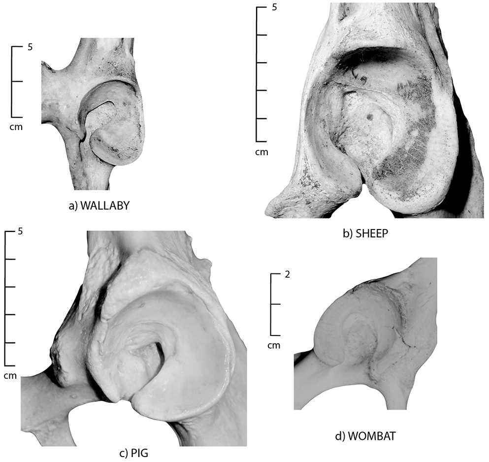
Figure 6.5: A detail image of an acetabulum; (a) wallaby, (b) sheep, (c) pig and (d) wombat; illustrating the lack of an acetabular notch, and the smooth, round shape.
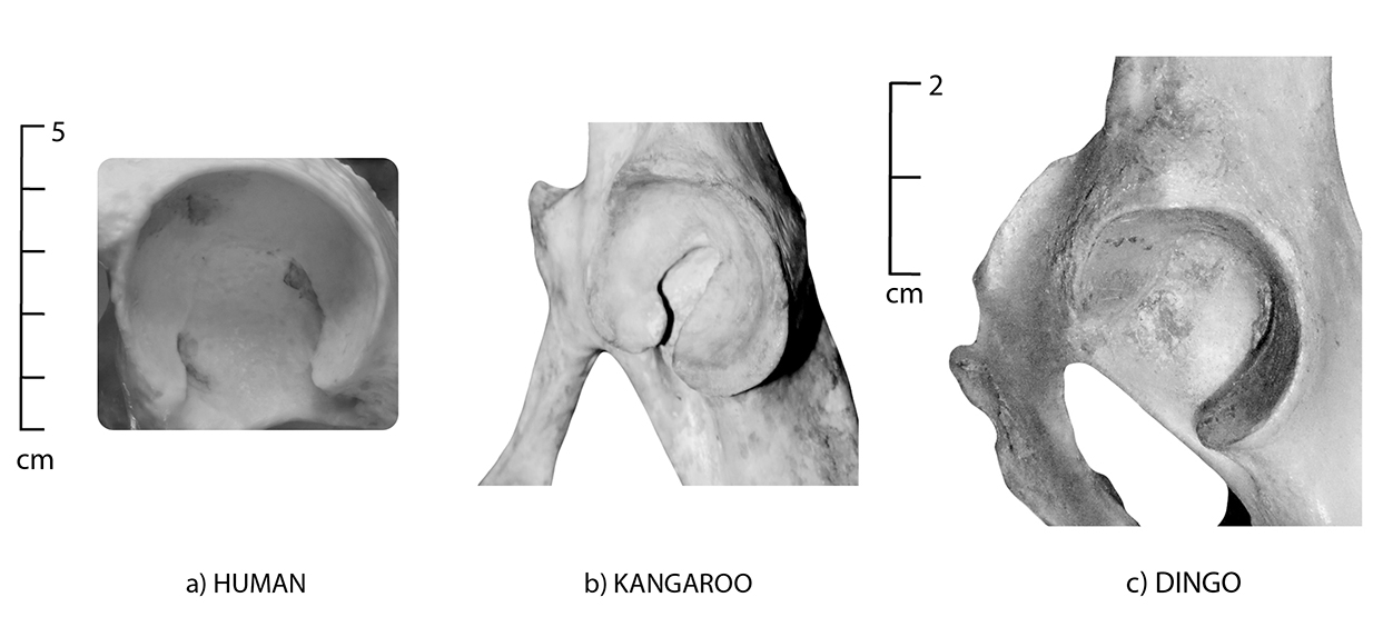
Figure 6.6: A detail image of an acetabulum; (a) human, (b) kangaroo and (c) dingo; illustrating a widely open acetabular notch (a) to an almost closed acetabular notch (b).
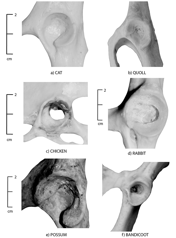
Figure 6.7: A detail image of (a) cat, (b) quoll, (c) chicken, (d) rabbit, (e) brushtail possum and (f) bandicoot acetabulum.
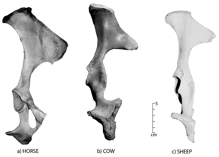
Figure 6.8: Innominate positioned to show the shape of the neck and blade; (a) kangaroo, (b) wallaby, (c) dingo, (d) cat, (e) quoll, (f) rabbit, (g) brushtail possum and (h) bandicoot. Note the long, thin tapering blade of the marsupials and the sickle-shaped hook of the wombat in Figure 6.10c. The dingo has a rounded blade.
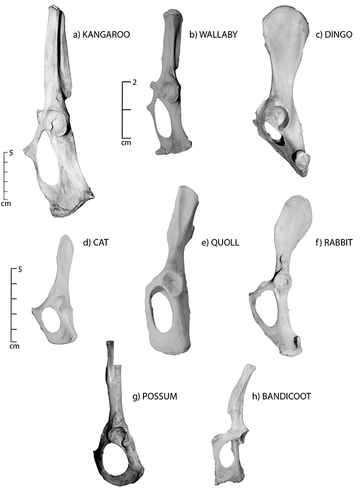
Figure 6.9: Pelvis positioned to show the morphology of the neck and the blade, especially the triangular crest of the ilium; (a) horse, (b) cow and (c) sheep.
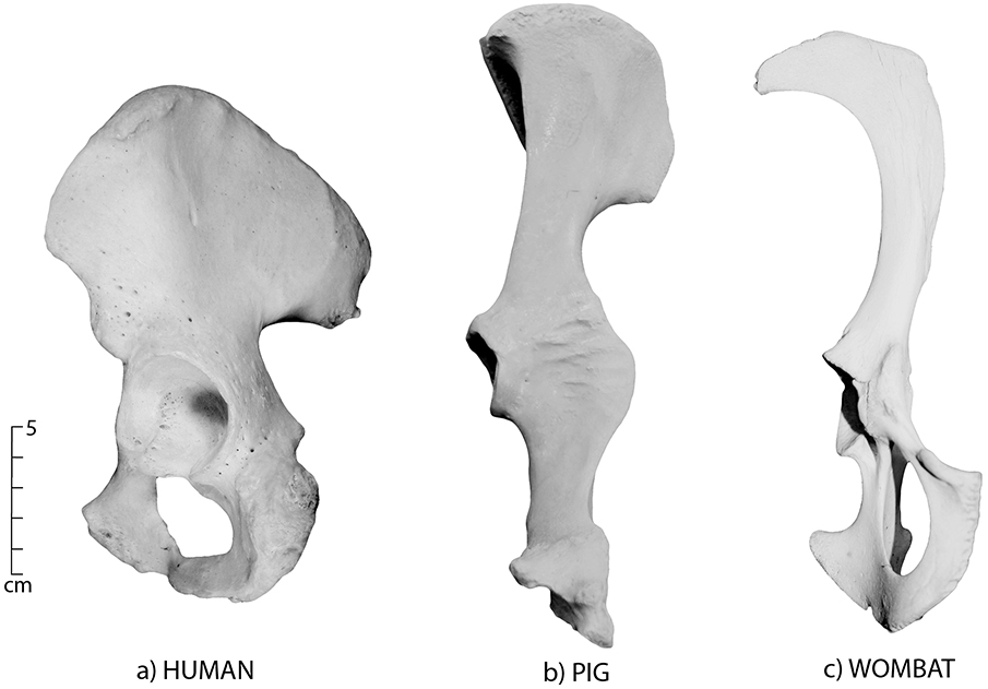
Figure 6.10: Pelvis positioned to show the morphology of the neck and the blade; (a) human, (b) pig and (c) wombat. The raised acetabulum of the pig is clear when compared to the shallower depression in the human.
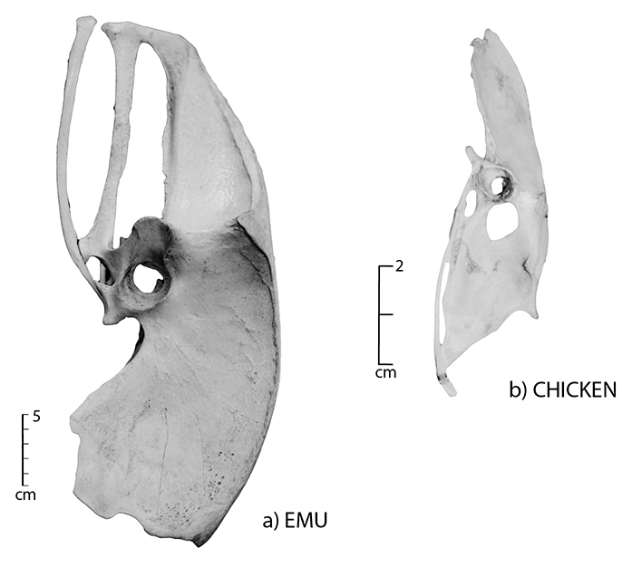
Figure 6.11: Articulated pelvis; (a) emu and (b) chicken.
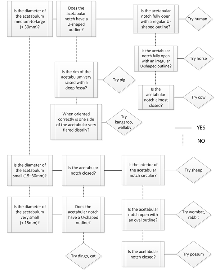 Figure 6.12: Pelvis decision process. Since the pelvis is generally fragmented in an archaeological assemblage, this decision process uses the morphological features of the acetabulum for species identification. |